文献:Au Nanocage Functionalized with Ultra-small Fe3O4 Nanoparticles for Targeting T1–T2Dual MRI and CT Imaging of Tumor
作者:Guannan Wang, Wei Gao, Xuanjun Zhang & Xifan Mei
文献链接:https://www.nature.com/articles/srep28258
摘要:
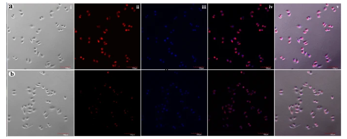
The targeting ability of F-AuNC@Fe3O4 to folate receptor-overexpressed cancer cells was studied using A549 cells as an example. To more directly display the targeting capabilities of the F-AuNC@Fe3O4, the fluorescence Cy5-PEG-SH was selected to label the nanoparticls. In order to give clear fluorescence imaging and retain the ability of bio-functionalization, firstly, the mixture of COOH-PEG-SH and fluorescence Cy5-PEG-SH with 9:1 ratio was used to functionalize the surface of AuNC@Fe3O4 and then fluorescent AuNC@Fe3O4 was conjugated with targeting molecular folic acid to form the fluorescent F-AuNC@Fe3O4 for targeting cancer cell. As a control group, the fluorescent AuNC@Fe3O4 without surface folic acid also was used, which showed similar physical properties to those of fluorescent F-AuNC@Fe3O4. Figure 4a,b row show the confocal images of A549 cells after incubation with fluorescent F-AuNC@Fe3O4 and AuNC@Fe3O4 suspensions at a concentration of 0.5 mg/mL for one hour, respectively. The fluorescence image shown in Fig. 4a(i) clearly shows the successful internalization of the fluorescent F-AuNC@Fe3O4 into A549 cells. The much brighter red emission observed in Fig. 4a(i) as compared to that in Fig. 4b(i) (A549 cell incubation with fluorescent AuNC@Fe3O4) indicates that more F-AuNC@Fe3O4 can be internalized into A549 cells via folate receptor-mediated endocytosis and the F-AuNC@Fe3O4 have more capability for cancer targeting recognition. These results confirm the F-AuNC@Fe3O4 can be effectively targeted to the folate receptor expressed cancer cells.

为了更直接地展示F-AuNC@Fe3O4,选择荧光Cy5-PEG-SH标记纳米颗粒。为了获得清晰的荧光成像并保持生物功能化的能力,首先,使用比例为9:1的COOH-PEG-SH和荧光Cy5-PEG-SH的混合物对表面进行功能化AuNC@Fe3O4然后是荧光AuNC@Fe3O4与靶向分子叶酸结合形成荧光F-AuNC@Fe3O4用于靶向*症细胞。
作为对照组,荧光AuNC@Fe3O4在没有表面叶酸的情况下,也使用了叶酸,其物理性质与荧光染料相似F-AuNC@Fe3O4.图4a、b行显示了A549细胞与荧光蛋白孵育后的共聚焦图像F-AuNC@Fe3O4和AuNC@Fe3O4浓度为0.5的悬浮液 分别以mg/mL的浓度持续1小时。图4a(i)所示的荧光图像清楚地显示了荧光的成功内化F-AuNC@Fe3O4进入A549细胞。
与图4b(i)相比,图4a(i)中观察到更亮的红色发射(A549细胞与荧光灯孵育AuNC@Fe3O4)表示更多F-AuNC@Fe3O4可通过叶酸受体介导的内吞作用内化到A549细胞中F-AuNC@Fe3O4具有更强的*症靶向识别能力。
相关推荐:
CY5-PEG-SH
mPEG-PEI-CY5
Cy7.5-PEG-AA
CY7.5-PEG-Biotin
CY7.5-PEG-DBCO
CY7.5-PEG-DSPE
CY7.5-PEG-FA
CY7.5-PEG-N3
CY7.5-PEG-NH2
CY7.5-PEG-SH
CY7-PEG-Biotin
CY7-PEG-DBCO
CY7-PEG-DMG
以上文章内容来源各类期刊或文献,如有侵权请联系我们删除!


 齐岳微信公众号
 官方微信
官方微信 库存查询
库存查询